Neuroradiology · Neuroanatomy · A teaching atlas
Learn to read
the brain.
CNS Atlas is a neuroradiology teaching atlas. Scroll CT, MRI, angiography and tractography in one registered space, with every structure, vessel and tract named and tied to what happens when it fails. Built by a neuroradiologist for residents, fellows and students.
One space. Every modality.CT, MRI, angiography and tractography, registered to the same 1 mm grid.
Named and explainedStructures, vessels and tracts carry clinical teaching cards.
Anatomy to findingGuided lessons link a lesion to its deficit.
The radiology atlas
Seven series.
Three overlays.
Pick a series and scroll it like a study. Turn on an overlay to name what you are looking at, or turn it off and read the images bare.
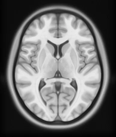 MRIT1ICBM 2009c template, 1 mm
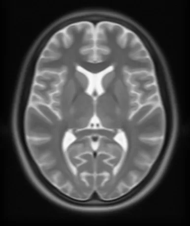 MRIT2Same template, CSF bright
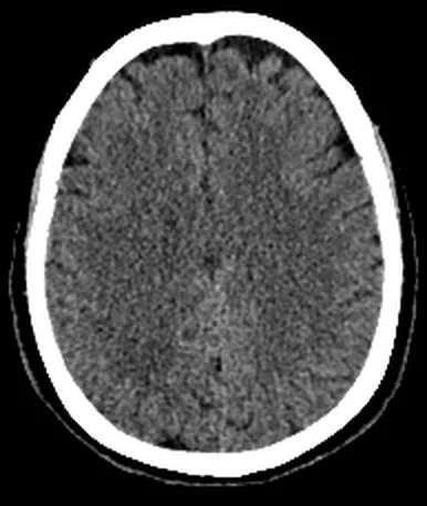 CTHead CTNon-contrast, brain window
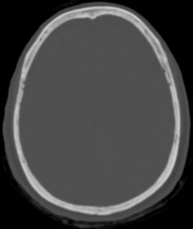 CTBoneSame scan, bone window
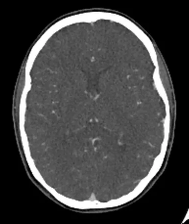 CTCTACT angiogram, vascular window
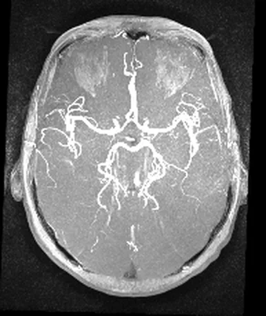 MRIMRATime-of-flight arteries
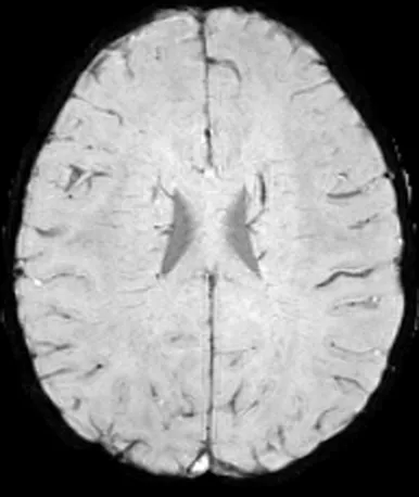 MRISWISusceptibility, veins dark
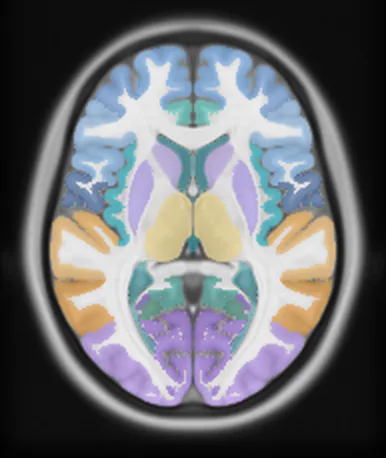 OverlayAnatomy51 structures with teaching cards
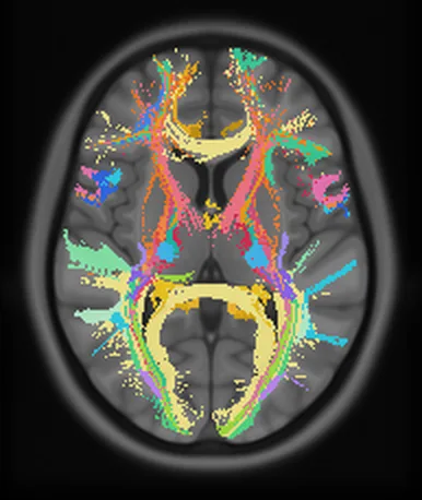 OverlayTracts46 bundles, HCP-1065 tractography
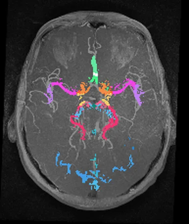 OverlayVessels23 named arteries, sinuses and veins
The curriculum
Begin with a question.
Follow the anatomy.
Each lesson starts with an anatomical relationship and builds toward understanding. Choose a pathway to study, or use the atlas to explore a structure in detail.
A way to study
Build understanding,
one relationship at a time.
The atlas is a companion for learning and teaching neuroanatomy. Use it alongside your course materials and the cited literature.
Explore the teaching atlas- Locate the structure
Orient yourself in the anatomy, then isolate a region or reveal deeper structures.
- Trace the relationship
Connect a structure to its pathways, functions, and relevant mechanisms.
- Apply and recall
Work through a guided example, check your understanding, and return to the anatomy.